- Background: Increased glycolytic activity is a hallmark of cancer, allowing staging and restaging with 18F-fluorodeoxyglucose-positron-emission-tomography (PET). Since interim-PET is an important prognostic tool in Hodgkin lymphoma (HL), the aim of this study was to investigate the expression of proteins involved in the regulation of glucose metabolism in the different HL subtypes and their impact on clinical outcome.
Methods: Lymph node biopsies from 54 HL cases and reactive lymphoid tissue were stained for glucose transporter 1 (GLUT1), lactate dehydrogenase A (LDHA) and lactate exporter proteins MCT1 and MCT4. In a second series, samples from additional 153 HL cases with available clinical data were stained for GLUT1 and LDHA.
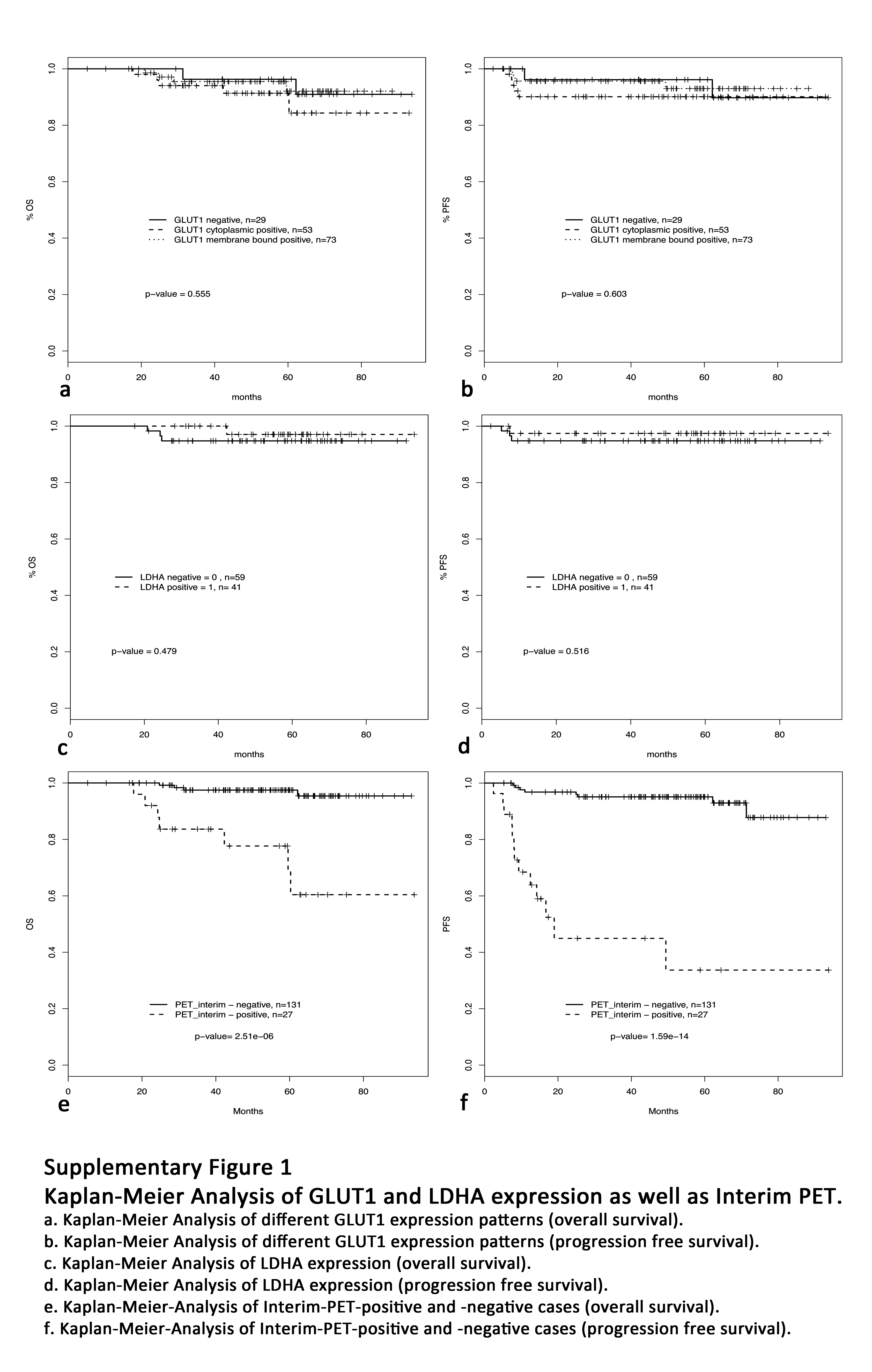
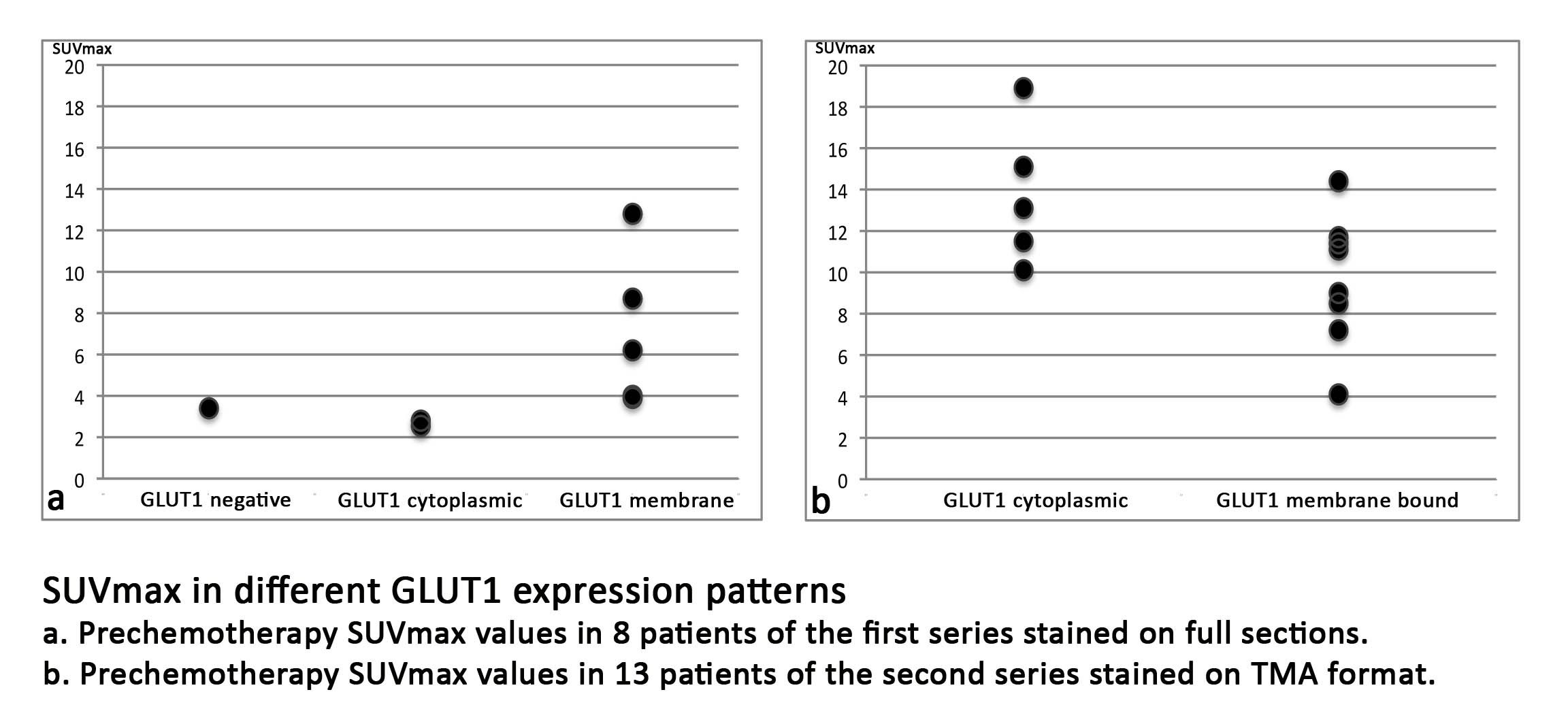
Results: Membrane bound GLUT1 expression was frequently observed in the tumor cells of HL (49% of all cases) but showed a broad variety between the different Hodgkin lymphoma subtypes: Nodular sclerosing HL subtype displayed a membrane bound GLUT1 expression in the Hodgkin-and Reed-Sternberg cells in 56% of the cases. However, membrane bound GLUT1 expression was more rarely observed in tumor cells of lymphocyte rich classical HL subtype (30%) or nodular lymphocyte predominant HL subtype (15%). Interestingly, in both of these lymphocyte rich HL subtypes as well as in progressively transformed germinal centers, reactive B cells displayed strong expression of GLUT1. LDHA, acting downstream of glycolysis, was also expressed in 44% of all cases. We evaluated the prognostic value of different GLUT1 and LDHA expression patterns; however, no significant differences in progression free or overall survival were found between patients exhibiting different GLUT1 or LDHA expression patterns. There was no correlation between GLUT1 expression in HRS cells and PET standard uptake values.
Conclusions: In a large number of cases, HRS cells in classical HL express high levels of GLUT1 and LDHA indicating glycolytic activity in the tumor cells. Although interim-PET is an important prognostic tool, a predictive value of GLUT1 or LDHA staining of the primary diagnostic biopsy could not be demonstrated. However, we observed GLUT1 expression in progressively transformed germinal centers and hyperplastic follicles, explaining false positive results in PET. Therefore, PET findings suggestive of HL relapse should always be confirmed by histology.
Metadaten| Author: | Sylvia HartmannORCiDGND, Claudio Agostinelli, Jürgen Diener, Claudia DöringGND, Stefano Fanti, Pier Luigi Zinzani, Andrea Gallamini, Lothar BergmannORCiDGND, Stefano Pileri, Martin-Leo HansmannGND |
|---|
| URN: | urn:nbn:de:hebis:30:3-277192 |
|---|
| DOI: | https://doi.org/10.1186/1471-2407-12-586 |
|---|
| ISSN: | 1471-2407 |
|---|
| Pubmed Id: | https://pubmed.ncbi.nlm.nih.gov/23228169 |
|---|
| Parent Title (English): | BMC cancer |
|---|
| Publisher: | BioMed Central |
|---|
| Place of publication: | London |
|---|
| Document Type: | Article |
|---|
| Language: | English |
|---|
| Date of Publication (online): | 2012/12/10 |
|---|
| Date of first Publication: | 2012/12/10 |
|---|
| Publishing Institution: | Universitätsbibliothek Johann Christian Senckenberg |
|---|
| Release Date: | 2013/01/17 |
|---|
| Tag: | GLUT1; Glycolysis; Hodgkin lymphoma; Warburg effect |
|---|
| Volume: | 12 |
|---|
| Issue: | 586 |
|---|
| Page Number: | 7 |
|---|
| Note: | © 2012 Hartmann et al.; licensee BioMed Central Ltd. This is an Open Access article distributed under the terms of the Creative Commons Attribution License ( http://creativecommons.org/licenses/by/2.0), which permits unrestricted use, distribution, and reproduction in any medium, provided the original work is properly cited. |
|---|
| HeBIS-PPN: | 319143988 |
|---|
| Institutes: | Medizin / Medizin |
|---|
| Dewey Decimal Classification: | 6 Technik, Medizin, angewandte Wissenschaften / 61 Medizin und Gesundheit / 610 Medizin und Gesundheit |
|---|
| Sammlungen: | Universitätspublikationen |
|---|
| Licence (German): |  Creative Commons - Namensnennung, Nicht kommerziell, Keine Bearbeitung 2.0 Creative Commons - Namensnennung, Nicht kommerziell, Keine Bearbeitung 2.0 |
|---|


 Creative Commons - Namensnennung, Nicht kommerziell, Keine Bearbeitung 2.0