Standardized mounting method of (zebrafish) embryos using a 3D-printed stamp for high-content, semi-automated confocal imaging
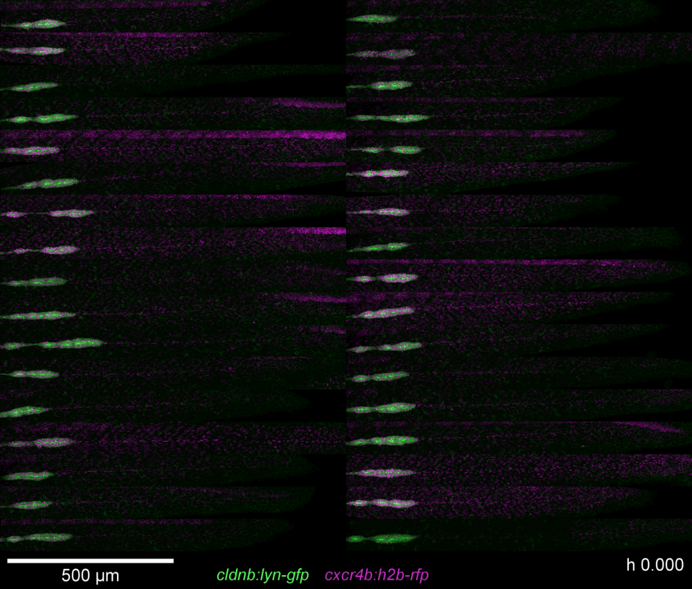
- Background: Developmental biology relies to a large extent on the observation and comparison of phenotypic traits through time using high resolution microscopes. In this context, transparent model organisms such as the zebrafish Danio rerio in which developing tissues and organs can be easily observed and imaged using fluorescent proteins have become very popular. One limiting factor however is the acquisition of a sufficient amount of data, in standardized and reproducible conditions, to allow robust quantitative analysis. One way to improve this is by developing mounting methods to increase the number of embryos that can be imaged simultaneously in near-to-identical orientation. Results: Here we present an improved mounting method allowing semi-automated and high-content imaging of zebrafish embryos. It is based on a 3D-printed stamp which is used to create a 2D coordinate system of multiple μ-wells in an agarose cast. Each μ-well models a negative of the average zebrafish embryo morphology between 22 and 96 h-post-fertilization. Due to this standardized and reproducible arrangement, it is possible to define a custom well plate in the respective imaging software that allows for a semi-automated imaging process. Furthermore, the improvement in Z-orientation significantly reduces post-processing and improves comparability of volumetric data while reducing light exposure and thus photo-bleaching and photo-toxicity, and improving signal-to-noise ratio (SNR). Conclusions: We present here a new method that allows to standardize and improve mounting and imaging of embryos. The 3D-printed stamp creates a 2D coordinate system of μ-wells in an agarose cast thus standardizing specimen mounting and allowing high-content imaging of up to 44 live or mounted zebrafish embryos simultaneously in a semi-automated, well-plate like manner on inverted confocal microscopes. In summary, image data quality and acquisition efficiency (amount of data per time) are significantly improved. The latter might also be crucial when using the services of a microscopy facility.
Download full text files
Additional Services


| Author: | David Kleinhans, Virginie LecaudeyORCiDGND |
|---|---|
| URN: | urn:nbn:de:hebis:30:3-519990 |
| DOI: | https://doi.org/10.1186/s12896-019-0558-y |
| ISSN: | 1472-6750 |
| Pubmed Id: | https://pubmed.ncbi.nlm.nih.gov/31640669 |
| Parent Title (English): | BMC biotechnology |
| Publisher: | BioMed Central |
| Place of publication: | London |
| Document Type: | Article |
| Language: | English |
| Year of Completion: | 2019 |
| Date of first Publication: | 2019/10/22 |
| Publishing Institution: | Universitätsbibliothek Johann Christian Senckenberg |
| Release Date: | 2019/12/16 |
| Tag: | 3D printed stamp; Automation; High content imaging; Multidimensional imaging; Quantitative imaging; Reproducibility; Standardization; Zebrafish |
| Volume: | 19 |
| Issue: | 1, Art. 68 |
| Page Number: | 10 |
| First Page: | 1 |
| Last Page: | 10 |
| Note: | Open Access: This article is distributed under the terms of the Creative Commons Attribution 4.0 International License (http://creativecommons.org/licenses/by/4.0/), which permits unrestricted use, distribution, and reproduction in any medium, provided you give appropriate credit to the original author(s) and the source, provide a link to the Creative Commons license, and indicate if changes were made. The Creative Commons Public Domain Dedication waiver (http://creativecommons.org/publicdomain/zero/1.0/) applies to the data made available in this article, unless otherwise stated. |
| HeBIS-PPN: | 459378406 |
| Institutes: | Biowissenschaften / Biowissenschaften |
| Dewey Decimal Classification: | 5 Naturwissenschaften und Mathematik / 59 Tiere (Zoologie) / 590 Tiere (Zoologie) |
| Sammlungen: | Universitätspublikationen |
| Open-Access-Publikationsfonds: | Biowissenschaften |
| Licence (German): |  Creative Commons - Namensnennung 4.0 Creative Commons - Namensnennung 4.0 |