- Background & Aims: Microvillus inclusion disease (MVID) is a congenital intestinal malabsorption disorder caused by defective apical vesicular transport. Existing cellular models do not fully recapitulate this heterogeneous pathology. The aim of this study was to characterize 3-dimensional intestinal organoids that continuously generate polarized absorptive cells as an accessible and relevant model to investigate MVID.
Methods: Intestinal organoids from Munc18-2/Stxbp2-null mice that are deficient for apical vesicular transport were subjected to enterocyte-specific differentiation protocols. Lentiviral rescue experiments were performed using human MUNC18-2 variants. Apical trafficking and microvillus formation were characterized by confocal and transmission electron microscopy. Spinning disc time-lapse microscopy was used to document the lifecycle of microvillus inclusions.
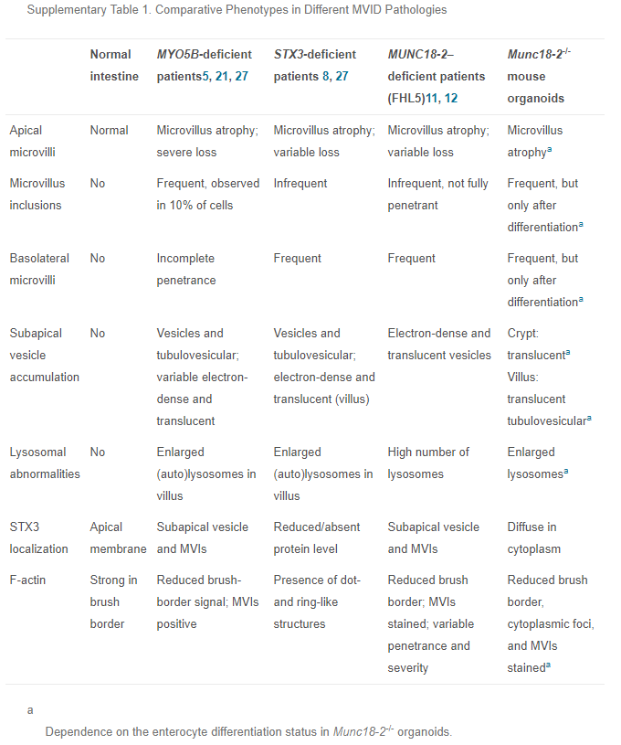
Results: Loss of Munc18-2/Stxbp2 recapitulated the pathologic features observed in patients with MUNC18-2 deficiency. The defects were fully restored by transgenic wild-type human MUNC18-2 protein, but not the patient variant (P477L). Importantly, we discovered that the MVID phenotype was correlated with the degree of enterocyte differentiation: secretory vesicles accumulated already in crypt progenitors, while differentiated enterocytes showed an apical tubulovesicular network and enlarged lysosomes. Upon prolonged enterocyte differentiation, cytoplasmic F-actin–positive foci were observed that further progressed into classic microvillus inclusions. Time-lapse microscopy showed their dynamic formation by intracellular maturation or invagination of the apical or basolateral plasma membrane.
Conclusions: We show that prolonged enterocyte-specific differentiation is required to recapitulate the entire spectrum of MVID. Primary organoids can provide a powerful model for this heterogeneous pathology. Formation of microvillus inclusions from multiple membrane sources showed an unexpected dynamic of the enterocyte brush border.
Metadaten| Verfasserangaben: | Mohammed Hossameldin MosaORCiDGND, Ophélie Nicolle, Sophia Maschalidi, Fernando E. Sepulveda, Aurelien Bidaud-Meynard, Constantin MencheORCiDGND, Birgitta E. MichelsORCiDGND, Grégoire Michaux, Geneviève de Saint Basile, Henner FarinORCiDGND |
|---|
| URN: | urn:nbn:de:hebis:30:3-479326 |
|---|
| DOI: | https://doi.org/10.1016/j.jcmgh.2018.08.001 |
|---|
| ISSN: | 2352-345X |
|---|
| Pubmed-Id: | https://pubmed.ncbi.nlm.nih.gov/30364784 |
|---|
| Titel des übergeordneten Werkes (Englisch): | Cellular and Molecular Gastroenterology and Hepatology |
|---|
| Verlag: | Elsevier |
|---|
| Verlagsort: | New York, NY |
|---|
| Dokumentart: | Wissenschaftlicher Artikel |
|---|
| Sprache: | Englisch |
|---|
| Jahr der Fertigstellung: | 2018 |
|---|
| Datum der Erstveröffentlichung: | 14.08.2018 |
|---|
| Veröffentlichende Institution: | Universitätsbibliothek Johann Christian Senckenberg |
|---|
| Datum der Freischaltung: | 08.11.2018 |
|---|
| Freies Schlagwort / Tag: | Apical Vesicular Transport; Brush Border Formation; Disease Modeling; Microvillus Atrophy |
|---|
| Jahrgang: | 6 |
|---|
| Ausgabe / Heft: | 4 |
|---|
| Seitenzahl: | 18 |
|---|
| Erste Seite: | 477 |
|---|
| Letzte Seite: | 493.e1 |
|---|
| Bemerkung: | © 2018 The Authors. Published by Elsevier Inc. on behalf of the AGA Institute. This is an open access article under the CC BY-NC-ND license (http://creativecommons.org/licenses/by-nc-nd/4.0/). |
|---|
| HeBIS-PPN: | 44009318X |
|---|
| Institute: | Biowissenschaften / Biowissenschaften |
|---|
| Angeschlossene und kooperierende Institutionen / Georg-Speyer-Haus |
|---|
| DDC-Klassifikation: | 5 Naturwissenschaften und Mathematik / 57 Biowissenschaften; Biologie / 570 Biowissenschaften; Biologie |
|---|
| Sammlungen: | Universitätspublikationen |
|---|
| Lizenz (Deutsch): |  Creative Commons - Namensnennung-Nicht kommerziell - Keine Bearbeitung 4.0 Creative Commons - Namensnennung-Nicht kommerziell - Keine Bearbeitung 4.0 |
|---|


 Creative Commons - Namensnennung-Nicht kommerziell - Keine Bearbeitung 4.0